Spatial multi-omics sequencing
Home
>
Sequencing
> Spatial multi-omics sequencing
Spatial Multi-Omics Sequencing Services — Map Gene Expression in Its Native Tissue Context
At N2Jenomics Lab Pvt. Ltd., we offer a comprehensive suite of Spatial Transcriptomics Sequencing Services that enables researchers to explore gene expression while preserving the spatial organization of tissues. Our advanced spatial multi-omics solutions combine state-of-the-art sequencing and imaging technologies, providing insights into cellular architecture, tissue heterogeneity, and molecular interactions that conventional transcriptomics cannot reveal.
Whether your study focuses on whole-transcriptome profiling in fresh frozen tissues or high-resolution targeted analysis in FFPE samples, our expert team delivers end-to-end support—from experimental design and platform selection to sequencing, bioinformatics, and biological interpretation.
Our Spatial Transcriptomics Platforms
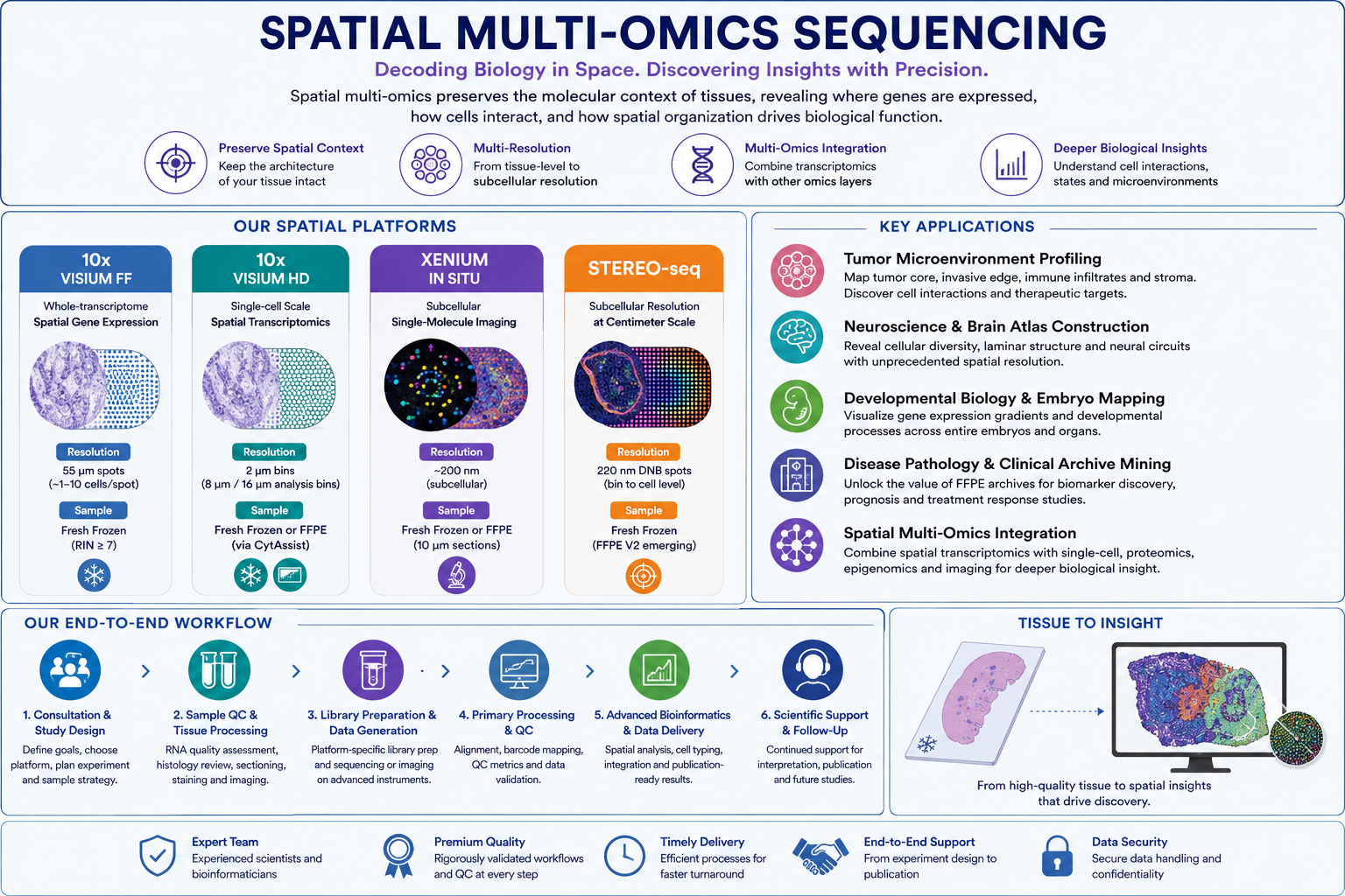
We provide four industry-leading spatial transcriptomics technologies to accommodate diverse research objectives:
• Visium HD™ for high-resolution spatial transcriptomics with enhanced cellular localization.
• Xenium™ In Situ for imaging-based, single-molecule RNA detection with subcellular precision.
• Stereo-seq for ultra-high-resolution spatial transcriptomics across large tissue sections.
High-Resolution Spatial Analysis
Our platform portfolio supports multiple levels of spatial resolution, allowing researchers to select the most appropriate technology for their study.
• 55 µm spot-based resolution for whole-transcriptome tissue profiling.
• Subcellular resolution down to approximately 200 nm for detailed cellular mapping.
• Single-molecule RNA detection for precise localization and quantification of individual transcripts.
Broad Sample Compatibility
Our workflows are optimized for a wide range of tissue preservation methods and biological samples, including:
• Fresh frozen tissue
• FFPE (Formalin-Fixed Paraffin-Embedded) tissue
• Fixed frozen tissue
We support research involving human, mouse, plant, animal, and other model or non-model organisms, depending on platform compatibility and project requirements.
N2Jenomics Lab Pvt. Ltd. provides a fully integrated service covering every stage of your spatial transcriptomics project, including:
• Scientific consultation and experimental design
• Tissue handling and quality assessment
• Sample preparation and optimization
• Spatial library preparation
• High-throughput sequencing or imaging
• Advanced bioinformatics analysis
• Cell type annotation and spatial mapping
• Differential gene expression analysis
• Pathway enrichment and biological interpretation
• Publication-ready figures, reports, and data deliverables
With cutting-edge technologies, experienced molecular biologists, and a dedicated bioinformatics team, N2Jenomics Lab Pvt. Ltd. empowers researchers to uncover spatial gene expression patterns, cellular interactions, and tissue microenvironments with exceptional precision, enabling breakthrough discoveries in cancer biology, neuroscience, immunology, developmental biology, pathology, and precision medicine.
Why Spatial Multi-Omics? Preserving the Molecular Architecture of Tissues
Traditional bulk RNA sequencing measures the average gene expression across millions of cells, masking the diversity and spatial organization within complex tissues. Although single-cell RNA sequencing (scRNA-seq) can identify individual cell populations, it requires tissue dissociation, resulting in the loss of critical spatial relationships between cells and their surrounding microenvironment.
Spatial transcriptomics overcomes these limitations by combining gene expression profiling with spatial localization, enabling researchers to visualize where genes are expressed while preserving the native architecture of the tissue. This provides a more complete understanding of cellular interactions, tissue organization, and biological function.
Recognized as Nature Methods' "Method of the Year" in 2020, spatial transcriptomics has rapidly evolved into a powerful suite of technologies that integrates sequencing, imaging, and multi-omics approaches. These platforms enable researchers to investigate complex biological systems with unprecedented spatial and molecular resolution.
At N2Jenomics Lab Pvt. Ltd., we offer a complete portfolio of industry-leading spatial transcriptomics platforms, allowing researchers to choose the most appropriate technology for their specific scientific objectives.
Our platform offerings include:
• Visium™ Spatial Gene Expression for unbiased whole-transcriptome profiling with spatial coordinates.
• Visium HD™ for enhanced spatial resolution and detailed tissue architecture analysis.
• Stereo-seq for ultra-high-resolution, genome-wide spatial transcriptomics across large tissue sections.
• Xenium™ In Situ for imaging-based, single-molecule RNA detection with true subcellular resolution and precise cell boundary mapping.
Tailored Solutions for Every Research Question
Each spatial technology provides unique strengths. Sequencing-based platforms generate comprehensive, genome-wide expression profiles while maintaining spatial context, making them ideal for tissue-wide discovery studies. Imaging-based approaches deliver highly sensitive visualization of selected transcripts at single-molecule resolution, enabling precise characterization of individual cells and their microenvironments.
By offering multiple complementary technologies under one roof, we help researchers select the most suitable platform based on sample type, resolution requirements, target coverage, and experimental goals. As projects progress, workflows can be seamlessly adapted or expanded without the need to transition to another service provider.
End-to-End Scientific Partnership
Beyond sequencing, our experienced scientists provide complete project support, including:
• Platform selection and feasibility assessment
• Experimental design and study planning
• Tissue processing and quality evaluation
• Library preparation or in situ assay workflows
• High-throughput sequencing or imaging
• Advanced spatial bioinformatics analysis
• Cell type annotation and spatial mapping
• Multi-omics data integration
• Biological interpretation and publication-ready reporting
With cutting-edge instrumentation, dedicated molecular biology expertise, and advanced bioinformatics capabilities, N2Jenomics Lab Pvt. Ltd. enables researchers to uncover the molecular geography of tissues, revealing how cells interact, organize, and function within their native biological environment across applications in cancer research, neuroscience, immunology, developmental biology, regenerative medicine, and precision healthcare.
Platform Comparison: Choosing the Right Spatial Technology
Each platform has distinct strengths. Use this table to match your experimental priorities to the right service — or consult our scientific team for a personalized recommendation.
At N2Jenomics Lab Pvt. Ltd., we offer a complete portfolio of industry-leading spatial transcriptomics technologies designed to address a broad range of biological and clinical research applications. Every project follows our comprehensive end-to-end workflow—from sample quality assessment and tissue processing to sequencing, advanced bioinformatics, and publication-ready data delivery.
Whether your goal is whole-transcriptome discovery, single-cell spatial profiling, subcellular transcript localization, or large-scale tissue mapping, our experts help you select the platform best suited to your research objectives.
- Integration with Visium™ datasets and single-cell RNA sequencing (scRNA-seq)
• Ideal Applications
- Biomarker validation
- Single-molecule RNA localization
- Cell morphology and transcript correlation
- Spatial immunology
- Clinical translational research
- Multiplex imaging workflows
Stereo-seq
Ultra-High-Resolution Spatial Transcriptomics for Large Tissue Sections
• Spatial Resolution
- Approximately 220 nm DNA Nanoball (DNB) capture spots, enabling subcellular spatial analysis across exceptionally large tissue areas.
• Transcript Coverage
- Whole-transcriptome profiling using:
- Poly(A)-based chemistry for fresh frozen tissues.
- Probe-based chemistry for emerging FFPE workflows.
• Supported Species
- Suitable for virtually any species with an available reference genome, including model and non-model organisms.
• Sample Compatibility
- Fresh frozen tissues (primary workflow).
- FFPE tissues through Stereo-seq V2 workflows.
• Bioinformatics Analysis
- SAW pipeline processing
- Large-scale tissue reconstruction
- Bin-to-cell aggregation
- Seurat and Scanpy analysis
- Cell clustering
- Spatial domain identification
- Cross-section tissue stitching
• Ideal Applications
- Whole-organ spatial mapping
- Whole-embryo transcriptomic atlases
- Brain and liver spatial studies
- Comparative genomics
- Cross-species spatial biology
- Large-area, high-resolution tissue profiling
End-to-End Spatial Transcriptomics Solutions
Regardless of the platform selected, every project at N2Jenomics Lab Pvt. Ltd. benefits from our integrated workflow, including:
- Scientific consultation and platform selection
- Experimental design and feasibility assessment
- Tissue processing and quality control
- Spatial library preparation or in situ assay setup
- High-throughput sequencing or imaging
- Comprehensive bioinformatics analysis
- Cell-type annotation and spatial mapping
- Multi-omics integration
- Biological interpretation
- Publication-ready reports, figures, and data deliverables
Our multidisciplinary team ensures that each project is optimized for your research objectives, delivering reliable, high-resolution spatial data that accelerates discoveries in cancer biology, neuroscience, immunology, developmental biology, regenerative medicine, pathology, and precision healthcare.
How to Choose: Decision Guide by Research Question
Match your primary research question to the platform best suited to answer it. Most complex projects benefit from a combination of platforms — our scientific team can help design a multi-platform strategy.
Research Question
Recommended Platform
Why
I want whole-transcriptome spatial expression in a non-human species
Visium FF
Only poly(A) capture method compatible with any reference genome; no species-specific probe design needed
I need single-cell-level spatial resolution from FFPE archives
Visium HD
2 µm bin size reaches single-cell scale with continuous coverage; CytAssist enables FFPE compatibility
I need exact subcellular transcript localization and cell morphology data
Xenium In Situ
Single-molecule FISH imaging at ~200 nm; DAPI-defined cell boundaries; highest spatial precision per transcript
I'm mapping an entire embryo or large organ section at high resolution
Stereo-seq
220 nm resolution + centimeter-scale capture area (up to 13 × 13 cm) — no other platform combines both
I want to discover novel spatial biology in a tumor microenvironment
Visium FF + scRNA-seq deconvolution
Unbiased whole-transcriptome spatial map, combined with matched scRNA-seq reference for cell-type resolution
I have a validated gene panel and need exact cell counts and morphology
Xenium In Situ
Targeted panel design; single-molecule sensitivity; integrates with DAPI, IF protein staining
I need a spatial atlas at single-cell scale with maximum transcriptome depth
Visium HD or Stereo-seq
Both offer near-single-cell resolution genome-wide; Visium HD is better for human/mouse FFPE; Stereo-seq for large-area or non-model applications
Key Applications of Spatial Multi-Omics
Spatial multi-omics is transforming biological and biomedical research by revealing where genes are expressed, how cells interact, and how tissue architecture influences biological function. By preserving spatial context, researchers can investigate complex cellular ecosystems that cannot be resolved using conventional bulk or single-cell sequencing alone.
At N2Jenomics Lab Pvt. Ltd., our comprehensive portfolio of Visium™, Visium HD™, Xenium™ In Situ, and Stereo-seq platforms enables researchers to select the most appropriate technology for virtually any spatial biology application.
1. Tumor Microenvironment (TME) Profiling
Cancer tissues consist of diverse cell populations that continuously communicate with one another. Spatial transcriptomics enables researchers to visualize the organization of tumor cells, immune infiltrates, stromal components, and surrounding healthy tissue within their native microenvironment.
Our spatial analysis workflows support:
• Tumor core and invasive margin characterization
• Immune cell infiltration mapping
• Cancer-associated fibroblast (CAF) analysis
• Spatial biomarker discovery
• Cell-cell communication and ligand-receptor interaction analysis
• Identification of therapeutic targets
• Precision oncology research
• Platform selection is optimized according to the desired spatial resolution, sample type, and research objectives.
2. Neuroscience & Brain Atlas Construction
The nervous system contains highly specialized cell populations arranged in complex anatomical structures. Spatial transcriptomics provides unprecedented insight into neuronal organization while preserving tissue architecture.
Applications include:
• Cortical layer characterization
• Hippocampal and cerebellar mapping
• Brain development studies
• Neurodegenerative disease research
• Neural circuit organization
• Cell-type-specific spatial expression profiling
• Construction of comprehensive brain atlases
By combining whole-transcriptome sequencing with single-cell and subcellular imaging technologies, researchers can generate highly detailed spatial maps of the nervous system.
3. Developmental Biology & Embryonic Research
Understanding embryonic development requires monitoring gene expression across both time and space. Spatial transcriptomics allows researchers to visualize developmental processes while maintaining the structural organization of tissues.
Common applications include:
• Whole-embryo spatial transcriptomics
• Organogenesis studies
• Cell lineage mapping
• Morphogen gradient analysis
• Tissue differentiation
• Developmental signaling pathways
• Comparative developmental genomics
• Large-area imaging platforms such as Stereo-seq enable high-resolution mapping across entire embryos and developing organs.
4. Disease Pathology & Clinical Research
Spatial technologies have significantly expanded the molecular analysis of archived clinical specimens, particularly FFPE (Formalin-Fixed Paraffin-Embedded) tissues.
Researchers can investigate:
• Disease-associated molecular signatures
• Biomarker discovery and validation
• Treatment response evaluation
• Tumor heterogeneity
• Tissue pathology
• Precision medicine research
• Retrospective clinical cohort studies
These capabilities allow valuable hospital and biobank specimens to be revisited using modern spatial genomics approaches.
5. Integrated Spatial Multi-Omics
Combining complementary technologies provides a more comprehensive understanding of complex biological systems.
Our integrated workflows can incorporate:
• Spatial transcriptomics
• Single-cell RNA sequencing (scRNA-seq)
• Bulk RNA sequencing
• Spatial proteomics
• Epigenomics
• Imaging data
• Histopathology
• Immunofluorescence
• Advanced bioinformatics pipelines integrate these datasets to identify cellular interactions, signaling pathways, and molecular mechanisms with greater biological accuracy.
What to Expect: Our End-to-End Spatial Transcriptomics Workflow
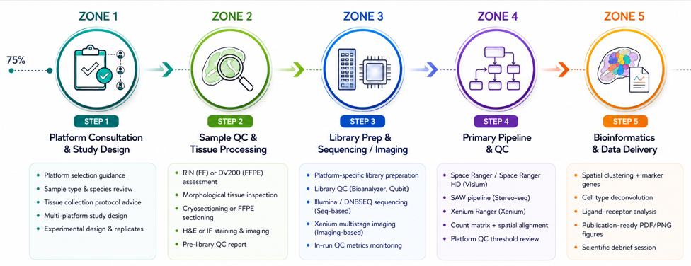
At N2Jenomics Lab Pvt. Ltd., every spatial transcriptomics project follows a carefully designed workflow that ensures high-quality data generation, rigorous quality control, and comprehensive scientific support from project initiation through publication.
All submitted specimens undergo comprehensive quality evaluation before processing.
Quality assessment includes:
• RNA integrity evaluation (RIN)
• FFPE quality assessment (DV200)
• Histological review
• Tissue morphology inspection
• Sample documentation
• Cryosectioning or FFPE section preparation
• H&E staining
• Immunofluorescence staining (when applicable)
If a sample does not meet quality requirements, our team provides a detailed evaluation and recommendations before proceeding.
Step 3. Library Preparation & Spatial Data Generation
Following successful quality control, platform-specific workflows are performed using validated laboratory protocols.
Depending on the selected technology, this stage includes:
• Spatial library preparation
• Probe hybridization
• Tissue imaging
• Barcode capture
• High-throughput sequencing
• In situ molecular imaging
• Library quality control
• Instrument performance validation
• Sequencing-based platforms utilize advanced Illumina or DNBSEQ systems, while imaging-based workflows employ dedicated high-resolution spatial imaging instruments.
Step 4. Primary Data Processing & Quality Control
Raw data are processed using platform-specific computational pipelines to generate accurate spatial gene expression datasets.
Primary analyses include:
• Image registration
• Sequence alignment
• Spatial barcode assignment
• Gene counting
• Cell segmentation
• Quality metric evaluation
• Technical performance assessment
• Every dataset undergoes stringent quality review before downstream biological analysis begins.
Step 5. Advanced Bioinformatics & Data Interpretation
Our experienced bioinformatics scientists perform comprehensive downstream analyses tailored to each project.
Available analyses include:
• Spatial clustering
• Differential gene expression
• Spatially variable gene identification
• Cell-type annotation
• Cell deconvolution
• Cell-cell communication analysis
• Ligand-receptor interaction analysis
• Pathway enrichment
• Spatial neighborhood analysis
• Multi-omics integration
• Interactive data visualization
• Final deliverables include publication-ready figures, comprehensive reports, processed datasets, and interactive visualization files compatible with leading spatial genomics software.
Step 6. Ongoing Scientific Collaboration
Our commitment extends beyond data delivery.
We provide continued scientific support through:
• Data interpretation consultations
• Follow-up analyses
• Manuscript preparation assistance
• Figure generation
• Reviewer response support
• Additional bioinformatics analyses
• Long-term collaboration for multi-phase projects
By combining advanced spatial transcriptomics technologies with experienced molecular biologists, bioinformaticians, and dedicated project managers, N2Jenomics Lab Pvt. Ltd. delivers complete spatial multi-omics solutions that help researchers uncover the spatial organization of biology and accelerate discoveries in oncology, neuroscience, immunology, developmental biology, pathology, regenerative medicine, and precision healthcare.
1. How do I choose the right spatial transcriptomics platform for my project?
Selecting the most suitable platform depends on your research objectives, sample type, desired spatial resolution, and the level of transcriptome coverage required.
Key considerations include:
Spatial Resolution:
• Visium™ Spatial Gene Expression provides multicellular resolution (55 µm spots), making it ideal for tissue-wide discovery studies.
• Visium HD™ offers near single-cell resolution with high-density capture features.
• Stereo-seq delivers ultra-high spatial resolution (approximately 220 nm) across exceptionally large tissue sections.
• Xenium™ In Situ enables single-molecule RNA detection with true subcellular precision.
Transcriptome Coverage:
• Visium™ and Stereo-seq perform unbiased whole-transcriptome profiling.
• Visium HD™ uses validated probe panels covering approximately 18,000 human and 20,000 mouse genes.
• Xenium™ targets predefined or custom gene panels for highly sensitive in situ analysis.
Sample Compatibility:
• Fresh frozen tissues are compatible with all major platforms.
• FFPE tissues are supported by Visium HD™ and Xenium™ workflows.
Species Support:
• Visium™ and Stereo-seq are suitable for many organisms with an available reference genome or transcriptome.
• Visium HD™ and Xenium™ are primarily optimized for human and mouse studies, with custom solutions available for selected applications.
Our scientific team will evaluate your samples and research goals to recommend the most appropriate platform—or a combination of platforms—for your study.
2. Can multiple spatial technologies be used within the same project?
Yes. Many advanced spatial biology studies combine complementary platforms to obtain a more comprehensive view of tissue organization and gene expression.
For example:
• Visium™ or Visium HD™ can be used for unbiased tissue-wide transcriptome profiling.
• Xenium™ In Situ can then validate and localize selected biomarkers at single-molecule resolution using adjacent tissue sections.
• Stereo-seq can provide ultra-high-resolution mapping across larger tissue areas.
Our team manages tissue sectioning, experimental coordination, and cross-platform bioinformatics integration to ensure consistent, biologically meaningful results.
3. What sample quality is recommended for spatial transcriptomics?
High-quality tissue preservation is essential for obtaining reliable spatial gene expression data.
• Fresh Frozen Samples
- Recommended RNA Integrity Number (RIN): ≥7
- Minimum acceptable RIN: ≥6
- Rapid tissue freezing and appropriate storage are strongly recommended.
• FFPE Samples
- Recommended DV200 value: ≥30%
- Proper fixation and processing improve RNA quality and downstream performance.
Before library preparation, we perform comprehensive tissue quality assessment and provide a detailed quality control report with recommendations regarding sample suitability.
4. Can spatial transcriptomics be performed on non-model organisms?
Yes. Several of our spatial transcriptomics platforms are well suited for non-model species.
Platforms such as Visium™ Spatial Gene Expression and Stereo-seq can be applied to a wide variety of organisms provided that suitable reference genome or transcriptome resources are available for downstream analysis.
These technologies have broad applications in:
• Agricultural research
• Plant genomics
• Aquatic biology
• Veterinary research
• Evolutionary biology
• Ecological genomics
• Comparative genomics
• For imaging-based platforms such as Visium HD™ and Xenium™ In Situ, species availability depends on validated or custom-designed probe panels. Our experts can advise on feasibility for your organism of interest.
5. What data and bioinformatics deliverables are included?
Each spatial multi-omics project includes comprehensive laboratory and bioinformatics deliverables designed to support downstream research and publication.
Typical deliverables include:
• Raw sequencing or imaging data
• Primary analysis outputs generated using platform-specific pipelines
• Sample quality control reports
• Spatial gene expression matrices
• Cell segmentation and annotation (where applicable)
• Spatial clustering analysis
• Differential gene expression analysis
• Spatially variable gene identification
• Cell-type deconvolution
• Cell–cell communication and ligand–receptor interaction analysis
• High-resolution visualization overlays on histological images
• Publication-ready figures and summary reports
• Interactive data formats compatible with leading spatial analysis software are also available to facilitate collaborative exploration and downstream interpretation.
6. Can you provide customized downstream bioinformatics analysis?
Absolutely. In addition to our standard analysis pipelines, we offer a wide range of advanced bioinformatics services tailored to your research objectives.
Customized analyses may include:
• Multi-platform spatial data integration
• Single-cell RNA-seq integration
• Spatial multi-omics integration
• Pathway and functional enrichment analysis
• Cell neighborhood analysis
• Spatial trajectory and lineage inference
• Biomarker discovery
• Comparative cohort analysis
• Customized statistical analysis
• Manuscript-ready visualizations and publication support
Our experienced bioinformatics team works closely with researchers to ensure that every dataset is analyzed using the most appropriate computational approaches for meaningful biological interpretation.
Address:Registered Office: 138, Patparganj Industrial Area, New Delhi – 110092, India
Operational Address: National Institute of Plant Genome Research (BRIC - NGGF)
Lab No. 206 and 207, Aruna Asaf Ali Marg, P.O. Box No. 10531,
New Delhi – 110067, India