Enzymatic Methylation Sequencing (EM-Seq) is an advanced DNA methylation analysis technology that provides accurate, genome-wide methylation profiling while preserving DNA integrity. Unlike traditional bisulfite sequencing, EM-Seq uses enzymatic conversion to distinguish methylated from unmethylated cytosines, minimizing DNA degradation and improving sequencing performance.
Its high sensitivity, low DNA input requirement, and excellent conversion efficiency make EM-Seq an ideal solution for precious clinical samples, low-input DNA, and high-quality epigenetic studies.
How EM-Seq Works
EM-Seq utilizes a series of enzymatic reactions to accurately identify methylated cytosines without exposing DNA to harsh chemical treatment.
The workflow involves:
• Protection of 5-methylcytosine (5mC) and 5-hydroxymethylcytosine (5hmC) through enzymatic oxidation.
• Enzymatic conversion of unmethylated cytosines into uracil.
• PCR amplification, where uracil is read as thymine while methylated cytosines remain unchanged.
• High-throughput sequencing and bioinformatics analysis to generate single-base resolution methylation profiles.
Because DNA damage is greatly reduced, EM-Seq produces higher-quality libraries and improved genome coverage compared with conventional bisulfite-based methods.
Why Choose EM-Seq?
Compared with traditional bisulfite sequencing, EM-Seq offers several important advantages:
• Preserves DNA integrity through gentle enzymatic conversion
• Single-base resolution DNA methylation profiling
• High conversion efficiency and methylation accuracy
• Low DNA input requirements
• Improved sequencing library complexity and genome coverage
• Compatible with standard next-generation sequencing platforms
• Supports both whole-genome and targeted methylation studies
• Ideal for low-quality or limited clinical samples, including circulating tumor DNA (ctDNA)
Applications
• Genome-Wide DNA Methylation Profiling
Generate comprehensive methylation maps for epigenetic research.
• Cancer Epigenetics
Identify methylation biomarkers associated with tumor development, progression, and therapeutic response.
• Biomarker Discovery
Discover and validate epigenetic biomarkers for disease diagnosis, prognosis, and precision medicine.
• Clinical Research
Analyze low-input and degraded DNA samples from clinical and liquid biopsy specimens.
• Developmental and Stem Cell Biology
Investigate epigenetic regulation during development, differentiation, and cellular reprogramming.
• Aging and Disease Research
Explore DNA methylation changes associated with aging, metabolic disorders, neurological diseases, and other complex conditions.
EM-Seq Workflow
Our optimized EM-Seq workflow ensures highly accurate and reproducible methylation analysis.
• Genomic DNA extraction and quality assessment
• Enzymatic protection of methylated cytosines
• Enzymatic conversion of unmethylated cytosines
• Library preparation and PCR amplification
• High-throughput sequencing
• Bioinformatics analysis, methylation calling, and comprehensive reporting
Key Benefits
Feature
EM-Seq
DNA Damage
Minimal
DNA Input Requirement
Low
Resolution
Single-base
Conversion Method
Enzymatic
Library Quality
High
Genome Coverage
Excellent
Suitable for Low-Input Samples
Yes
Clinical Sample Compatibility
Excellent
EM-Seq combines high sensitivity, exceptional accuracy, and superior DNA preservation, making it an excellent choice for modern epigenetic research and next-generation DNA methylation analysis.
Advantages of EM-Seq
EM-Seq offers significant improvements over conventional bisulfite sequencing by preserving DNA integrity while delivering highly accurate methylation profiling.
• Low DNA Input Requirements
Produces reliable methylation data from minimal DNA quantities, making it ideal for precious, degraded, or low-input samples such as circulating tumor DNA (ctDNA), FFPE tissues, and limited clinical specimens.
• Superior DNA Preservation
Gentle enzymatic conversion minimizes DNA fragmentation, resulting in higher-quality libraries and improved sequencing performance compared with bisulfite-based methods.
• Enhanced Genome Coverage
Generates longer DNA fragments with more uniform genome representation, reducing sequencing gaps and improving coverage across challenging genomic regions.
• Improved GC-Rich Region Analysis
Provides balanced coverage across both GC-rich and AT-rich regions, enabling more accurate methylation analysis of CpG islands and regulatory elements.
• High Sensitivity and Accuracy
Delivers single-base resolution methylation detection with excellent conversion efficiency, allowing precise identification of methylated cytosines throughout the genome.
• Greater Biomarker Discovery Potential
Improved CpG island coverage increases the likelihood of detecting biologically relevant methylation changes associated with disease development and progression.
Applications of EM-Seq
• Cancer Research and Liquid Biopsy
Identify methylation biomarkers from circulating cell-free DNA (cfDNA) for early cancer detection, disease monitoring, and precision oncology.
• Epigenetics Research
Investigate genome-wide DNA methylation patterns to understand gene regulation, cellular differentiation, and epigenetic mechanisms.
• Low-Input Sample Analysis
Generate high-quality methylation profiles from limited or precious samples, including embryos, stem cells, reproductive cells, and other scarce biological materials.
• Biomarker Discovery and Validation
Discover and validate DNA methylation biomarkers for disease diagnosis, prognosis, and therapeutic response.
• Disease Mechanism Studies
Explore epigenetic alterations associated with cancer, neurological disorders, autoimmune diseases, aging, and other complex conditions.
• Drug Discovery and Development
Assess epigenetic responses to therapeutic compounds and identify methylation-based biomarkers for drug efficacy and safety evaluation.
• Evolutionary and Agricultural Research
Compare DNA methylation patterns across species, populations, and breeding lines to investigate adaptation, evolution, and trait regulation.
• Transcription Start Site (TSS) Methylation Analysis
Accurately profile methylation within promoter regions and transcription start sites to better understand transcriptional regulation and gene expression.
DNA Methylation Analysis Platform Comparison
Technology
Resolution
Typical Data Output
CpG Coverage
Recommended DNA Input
Conversion Method
Best Suited For
850K Methylation Array
Single-base
Array-based
~850,000 CpG sites
≥250 ng
Bisulfite Conversion
Large cohort studies and clinical methylation profiling
Whole Genome Bisulfite Sequencing (WGBS)
Single-base
~90 Gb
~5 million CpG sites
≥1 µg
Bisulfite Conversion
Comprehensive whole-genome methylation analysis
Methyl Capture Sequencing (MC-Seq)
Single-base
~20 Gb
~2.7 million CpG sites
≥1 µg
Bisulfite Conversion
Targeted methylome profiling with reduced sequencing cost
Enzymatic Methylation Sequencing (EM-Seq)
Single-base
~25 Gb
~4 million CpG sites
≥10 ng
Enzymatic Conversion
Low-input samples and high-quality genome-wide methylation analysis
Single-Cell WGBS (scWGBS)
Single-base
~15 Gb
~5 million CpG sites
≥10 ng*
Bisulfite Conversion
Single-cell epigenomics and cellular heterogeneity studies
Pyrosequencing
Single-base
Targeted (50–90 bp)
Target-specific
≥500 ng
Bisulfite Conversion
Targeted methylation validation and locus-specific analysis
*Actual DNA input requirements may vary depending on sample type, library preparation strategy, and project design.
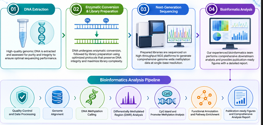
EM-Seq Workflow
Our standardized EM-Seq workflow is designed to deliver highly accurate, reproducible, and comprehensive DNA methylation profiling.
Step 1. DNA Extraction
High-quality genomic DNA is extracted and assessed for purity and integrity to ensure optimal sequencing performance.
DNA undergoes enzymatic conversion, followed by library preparation using optimized protocols that preserve DNA integrity and maximize library complexity.
Step 3. Next-Generation Sequencing
Prepared libraries are sequenced on high-throughput NGS platforms to generate comprehensive genome-wide methylation data at single-base resolution.
Step 4. Bioinformatics Analysis
Our experienced bioinformatics team performs comprehensive downstream analysis, including:
• Quality control and data processing
• Genome alignment
• DNA methylation calling
• Differentially methylated region (DMR) analysis
• CpG island and promoter methylation analysis
• Functional annotation and pathway enrichment
• Publication-ready figures and a comprehensive analysis report
Service Specification
Sample Requirements
Sample Types: Tissues, cells, and body fluids of cancer patients. Among the body fluid samples, plasma is recommended over serum to minimize interference from cfDNA derived from lymphocytes.
Note: Sample amounts are listed for reference only. For detailed information, please contact us with your customized requests.
Sequencing Strategies
Platform: High-throughput sequencing platforms such as Illumina HiSeq series.
Depth of coverage: ≥ 30x.
Quality score: More than 80% of bases with a ≥Q30 quality score.
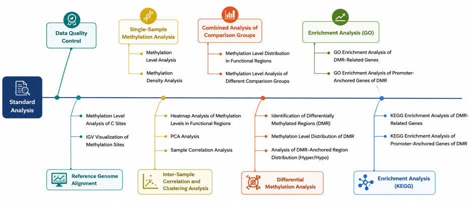
Bioinformatics Analysis We provide multiple customized bioinformatics analyses: 1. Genome Alignment Statistics 2. Overall Methylation Level Assessment
2.1 CpG Methylation Level Distribution
2.2 CpG Methylation Coverage
3. Methylation Site Statistics and Inter-sample Comparison
3.1 Methylation Level Correlation Analysis
3.2 Principal Component Analysis (PCA) of Methylation Levels
3.3 Cluster Analysis of Methylation Levels
4. Differential Methylation Site Analysis
4.1 Annotation of Differential Methylation Sites
4.2 Visualization of Differential Methylation Sites
4.3 Enrichment Analysis of Differential Methylation Sites
5. Differential Methylation Region Analysis
5.1 Annotation of Differential Methylation Sites
5.2 Visualization of Differential Methylation Sites
5.3 Enrichment Analysis of Differential Methylation Sites
Note: Recommended data outputs and analysis contents displayed are for reference only. For detailed information, please contact us with your customized requests.
Analysis Pipeline
Deliverables
• Original Sequencing Data
• Experimental Results
• Data Analysis Report
• Detailed Methylation Profiling
1. What are the sample requirements for genomic DNA (gDNA) and cell-free DNA (cfDNA)?
For optimal EM-Seq performance, high-quality DNA is recommended.
Recommended input:
• Genomic DNA (gDNA): ≥20 ng total DNA, concentration ≥2 ng/µL
• Cell-free DNA (cfDNA): ≥20 ng total DNA, concentration ≥2 ng/µL
To ensure efficient enzymatic conversion, DNA should be free from contaminants. Avoid using EDTA or EB buffer as the final solvent, as these may reduce enzyme performance and affect sequencing quality.
2. What precautions should be taken when preparing serum or plasma samples?
Proper sample handling is essential for obtaining high-quality cfDNA.
Recommended practices include:
• Process whole blood as soon as possible after collection.
• Separate serum or plasma promptly to minimize DNA degradation.
• Store serum or plasma at −80°C for long-term preservation.
• Avoid repeated freeze–thaw cycles, which can compromise sample integrity.
• Do not freeze whole blood before plasma or serum separation.
Following these guidelines helps maximize DNA quality and improves downstream EM-Seq performance.
3. How is the enzymatic conversion efficiency evaluated?
EM-Seq determines methylation by converting unmethylated cytosines while preserving methylated cytosines. Since the methylation status of experimental samples is unknown, conversion efficiency cannot be measured directly from the sample DNA.
Instead, unmethylated lambda DNA is included as an internal negative control during library preparation. Because all cytosines in lambda DNA are expected to undergo enzymatic conversion, the measured conversion rate of this control provides a reliable estimate of the overall enzymatic conversion efficiency for the experiment.
Address:Registered Office: 138, Patparganj Industrial Area, New Delhi – 110092, India
Operational Address: National Institute of Plant Genome Research (BRIC - NGGF)
Lab No. 206 and 207, Aruna Asaf Ali Marg, P.O. Box No. 10531,
New Delhi – 110067, India